Equine Distal Limb & Foot Instructional Models
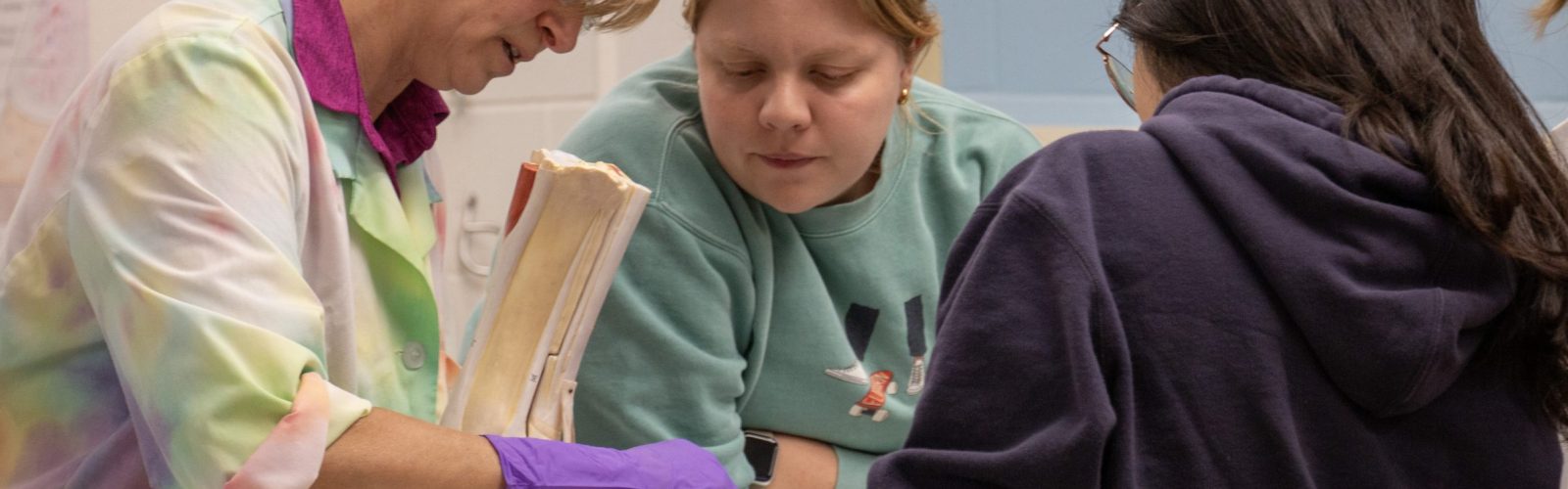
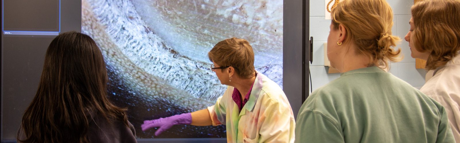
High-fidelity equine models created to reinforce the complex anatomy of the hoof and distal limb, including the dermal components, tendons, pulse points and nerve blocks. Related supportive content was created for use during active learning labs.
Original active learning exercises (ALEs) documents for the equine foot are provided, as well as four new ALEs for the distal limb model. Included also is our community agreement.
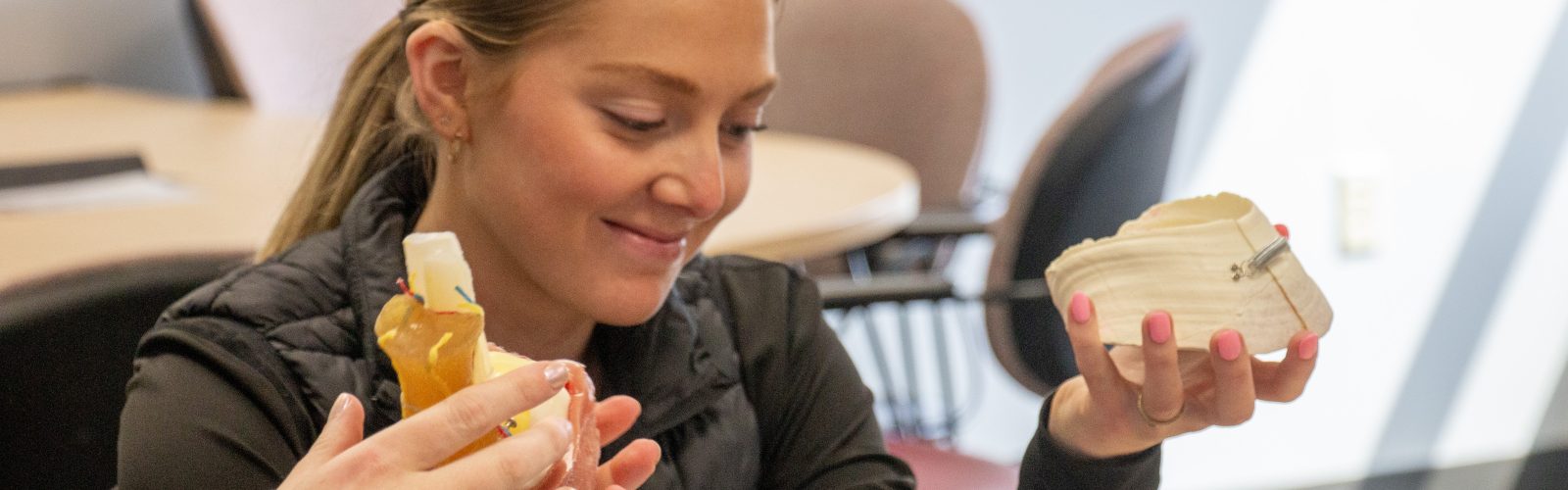
Equine Foot Instructional Model
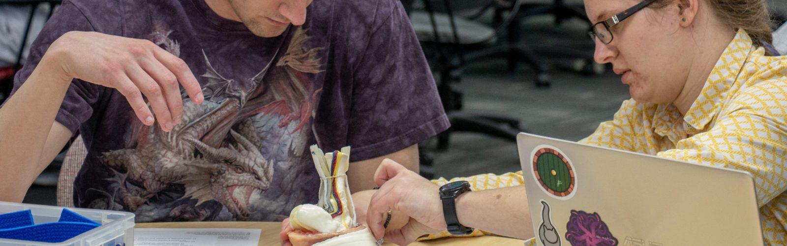
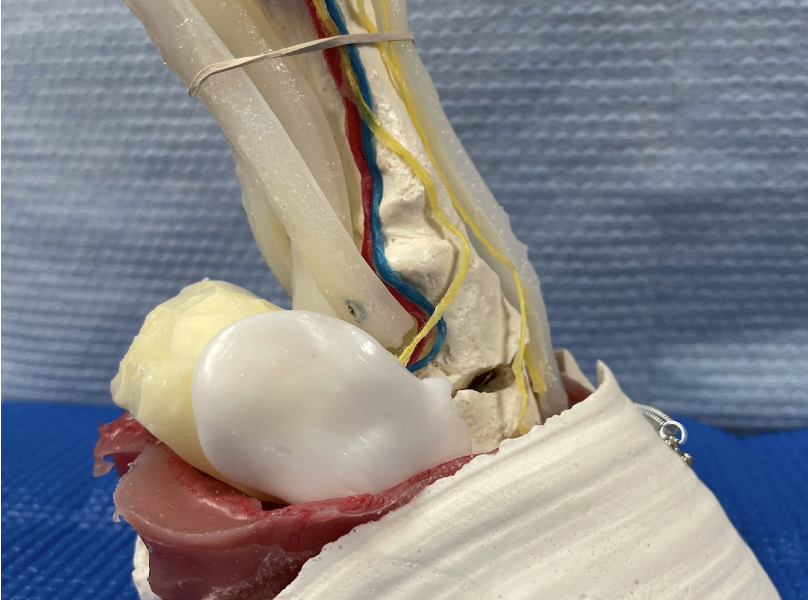
This equine foot model was the original creation, and enables users to study the complex structural and functional components found within the equine foot. There is a video describing its components in detail.
This version was created using a combination of resin, silicone, shapeable plastic, waxed string, red-dyed gauze, and a few small eyelets.
This version is very effective at teaching multiple concepts, but lacks moveable joints and contains only very distal structures. Hence the development of our more advanced model.
This is an accordion element with a series of buttons that open and close related content panels.
Active Learning Exercises for the Equine Foot Model
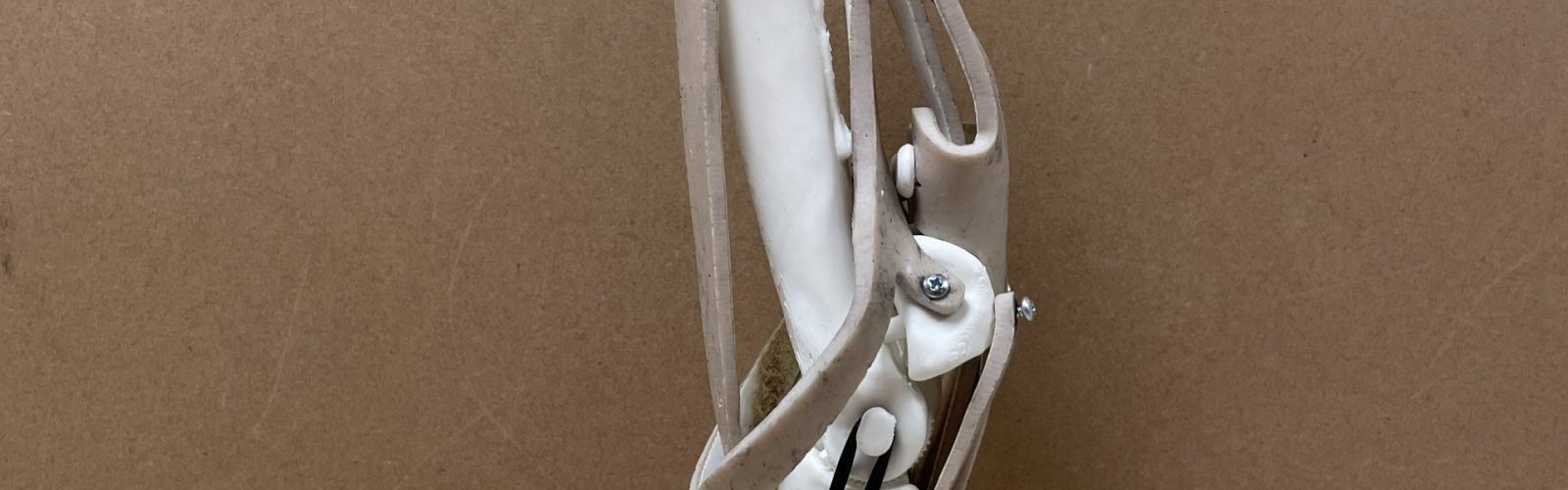
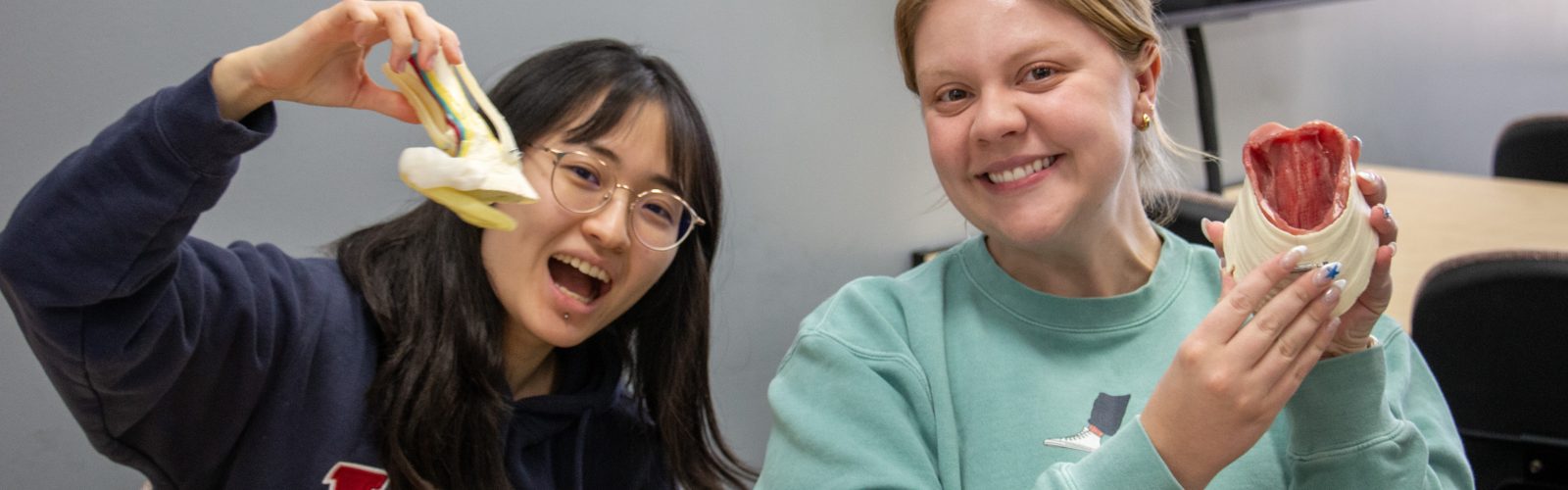
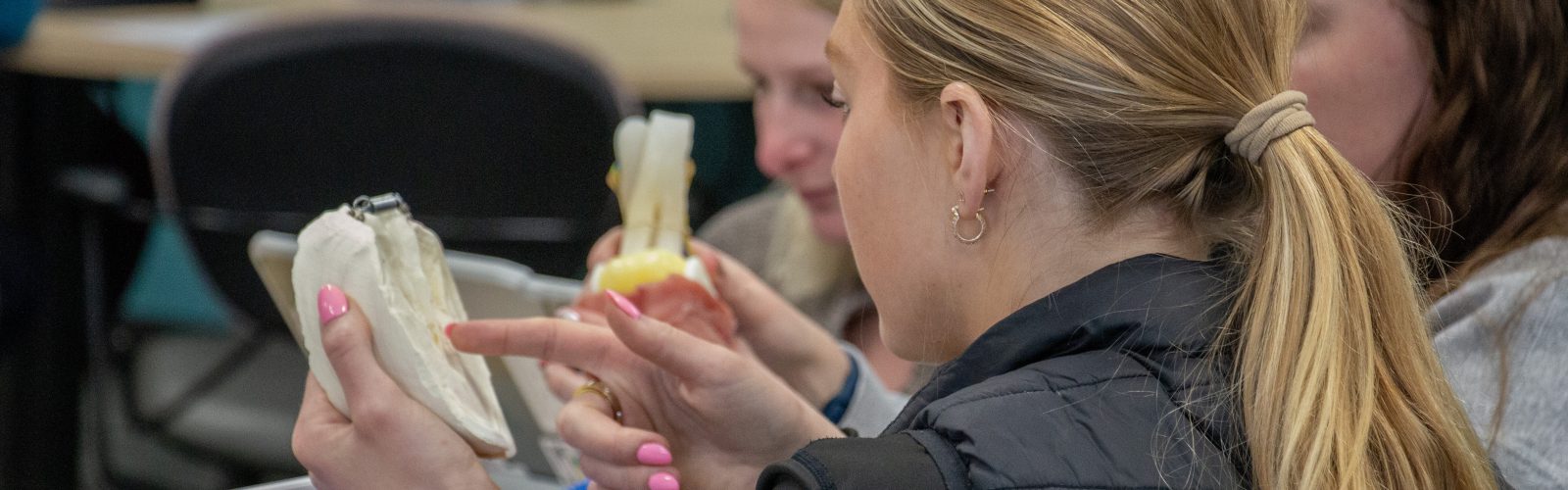
Equine Distal Limb Instructional Model
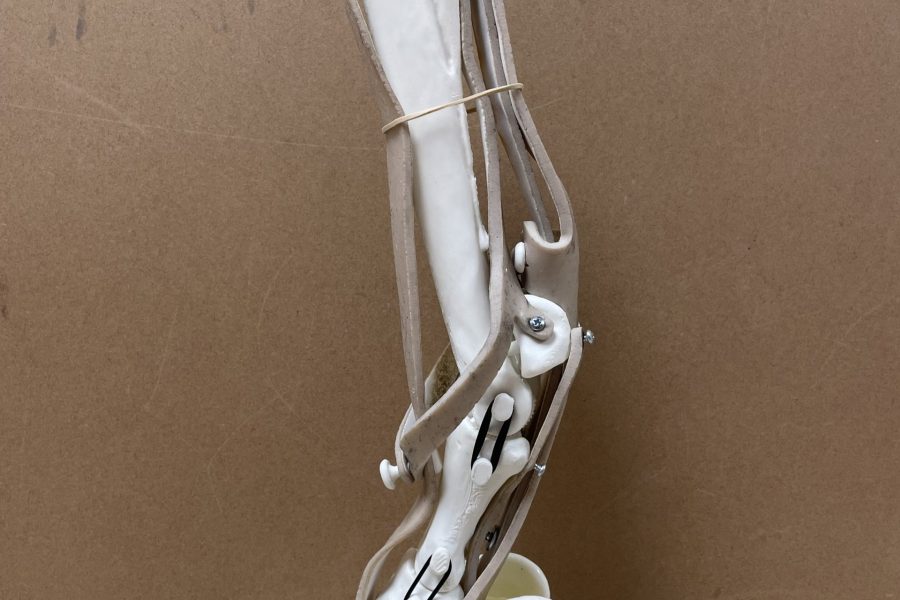
Our newest version consists of 3D printed bones, including the cannon bone, splint bones, long and short pastern bones, and the coffin bone, plus all distal sesamoids. This model has fully articulating joints with a ratchet mechanism to hold it in flexed or extended positions for studying. As it includes the entire distal limb, it also simulates more tendinous and ligamentous supportive structures, such as the interosseous ligament by use of reinforced laser-cut silicone sheets.
This model uses the same foot components as the original model to allow study of the dermal and epidermal components of the equine foot, as well.
This is an accordion element with a series of buttons that open and close related content panels.
Active Learning Exercises for the Equine Distal Limb Model
"Failure happens all the time. It happens every day in practice. What makes you better is how you react to it."
Mia Hamm (professional soccer player, olympian, writer)