Building the Female Urogenital Tract Model
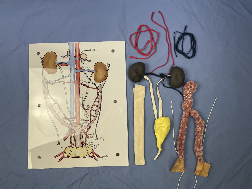
Deconstructed Model
These are the basic components of building the female urogenital tract model. Photo by McLean Gunderson.
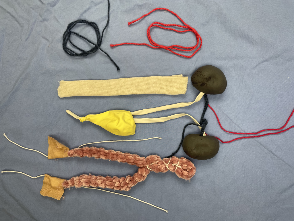
Model Components
These components represent the various organs and structures assembled during use of this model. Photo by McLean Gunderson.
Building the female urogenital tract is completed in our small animal anatomy course as an active learning exercise (ALE). It is one of many active learning exercises that stress the topographic relationships and vasculature of the pelvic and abdominal cavities.
This model has a plexiglass base with an image sandwiched in between. Press’n Seal® is used to recreate the serous membranes and secure items anatomically in relation to the board and each other. Colored string is used to outline arterial and venous blood flow. The remaining urogenital tract organs and structures are added in. Construction materials consist of string. thick yarn, balloons, and (dried) modeling foam. The distal termination of the colon/rectum are also included as a piece of stockinette to complete the organs of the pelvic cavity.
All but the Press’n Seal® is reusable. Included below are video instructions on the build, the Active learning exercise we use, as well as the base image.
“Optimism is the faith that leads to achievement. Nothing can be done without hope and confidence.”