Blood Flow Through the Uterus Model
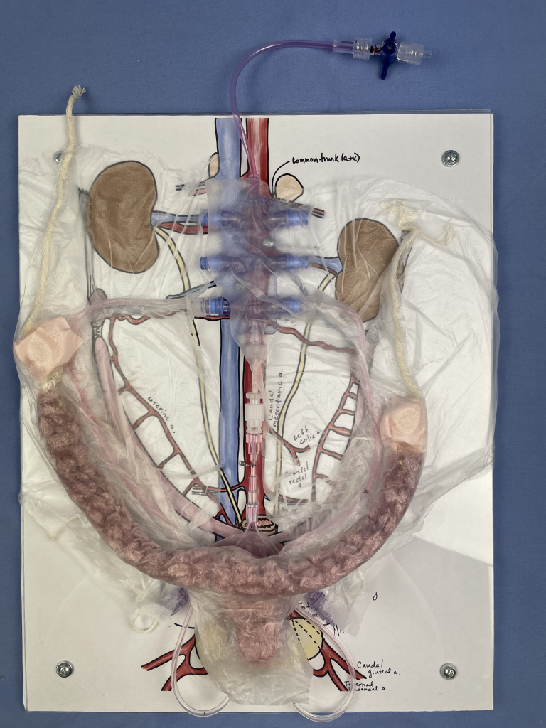
Completed Blood Flow Mode
Red colored fluid circulates via syringe. Broad, round and suspensory ligaments are featured. Photo by McLean Gunderson.
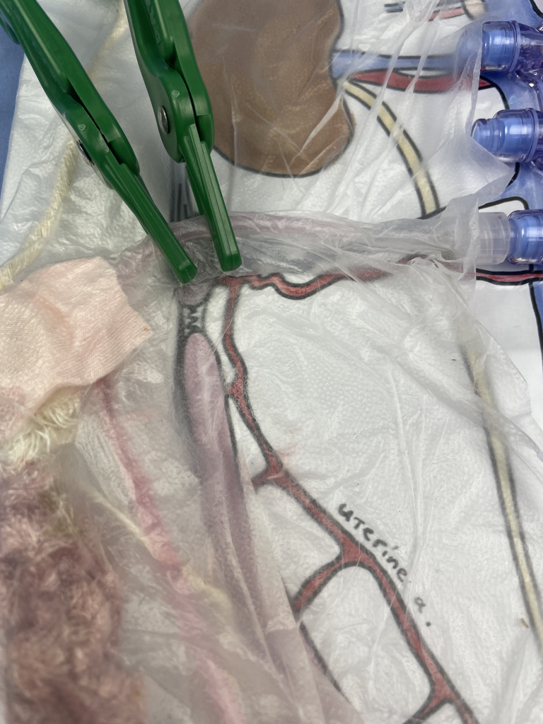
Clamps on Ovarian Pedicle
Hemostasis is accomplished through application of vascular clamps.
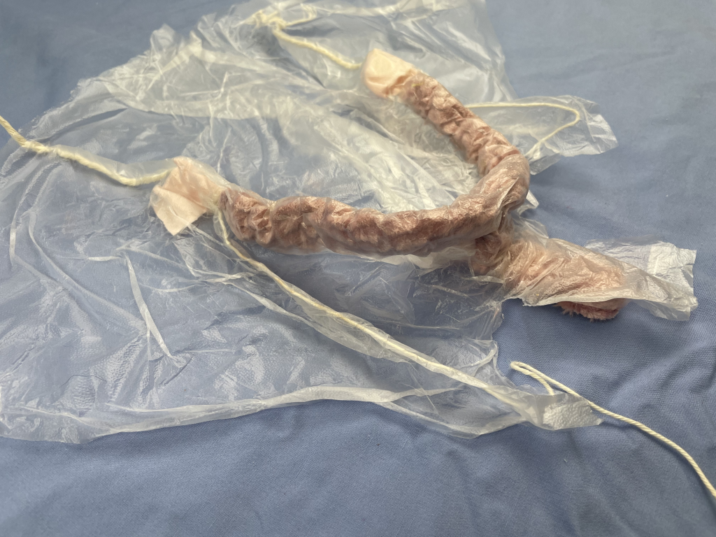
Featured Ligaments
Model has round, suspensory and broad ligaments. Blood flow is also in anatomically correct position. Photo by McLean Gunderson.
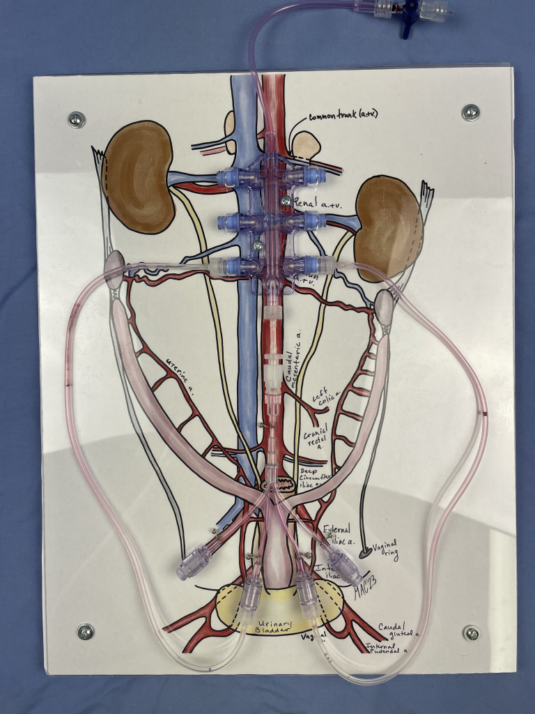
Collateral Circulation
Collateral circulation of the uterus & ovaries shown via red colored fluid pushed through the model. Photo by McLean Gunderson.
This model is used in an active learning activity for small animal anatomy. It is used to reinforce the anatomy and topography of the female reproductive tract in a clinically relevant context- an ovariohysterectomy (OHE)/spay surgical procedure.
Red fluid pushed by syringe through tubing mimics the collateral circulation servicing the uterus and ovaries in the cat and dog. String simulates the suspensory and round ligaments. Press’NSeal® secures the uterus, ovaries, vasculature, suspensory, and round ligaments within the broad ligament and secures it to the base in an anatomically correct position. Students apply plastic clamps to provide hemostasis at the ovarian pedicle and at the uterine body, as would be done in a spay procedure.
Ligatures can be applied and hemostasis checked when teaching more advanced clinical skills.
“Life is not easy for any of us. But what of that? We must have perseverance and above all confidence in ourselves. We must believe that we are gifted for something and that this thing must be attained.”